Program Results
2023 Annual report of Yushan Young scholar Prof Kuan-Ju Lu
Introduction to the event
A. Using dyes, fluorescent proteins, and gene guns, we tested the characteristics of protein transport
in Arabidopsis. Surprisingly, the protein transport in Arabidopsis was found to be less efficient than
predicted. We are currently actively investigating the regulatory mechanisms of protein transport
through the native cytoplasmic streaming and their similarity to known phenomena in vascular plants.
B. To understand how viruses utilize the native cytoplasmic streaming for movement, we established
a biotin labeling system. With this system, we have isolated approximately 200 target genes that may
be associated with viral movement proteins. Currently, we are testing the positive correlation with
viral movement for five high-potential genes and obtain one promising candidate gene that affects
the movement of virus. We will soon proceed to further understand the mechanism behind the
interference.
C. To gain further insights into the possible mechanisms influencing viral movement around the native
cytoplasmic streaming, we set up a more advanced native cytoplasmic streaming separation system.
This system is now combined with the biotin labeling system, and we are currently analyzing the genes
that might be involved in viral protein movement within the native cytoplasmic streaming.
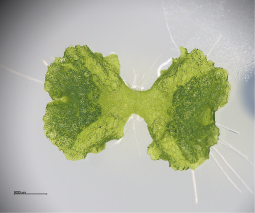
Fig 1: A 10-day-old Marchantia polymorpha plant
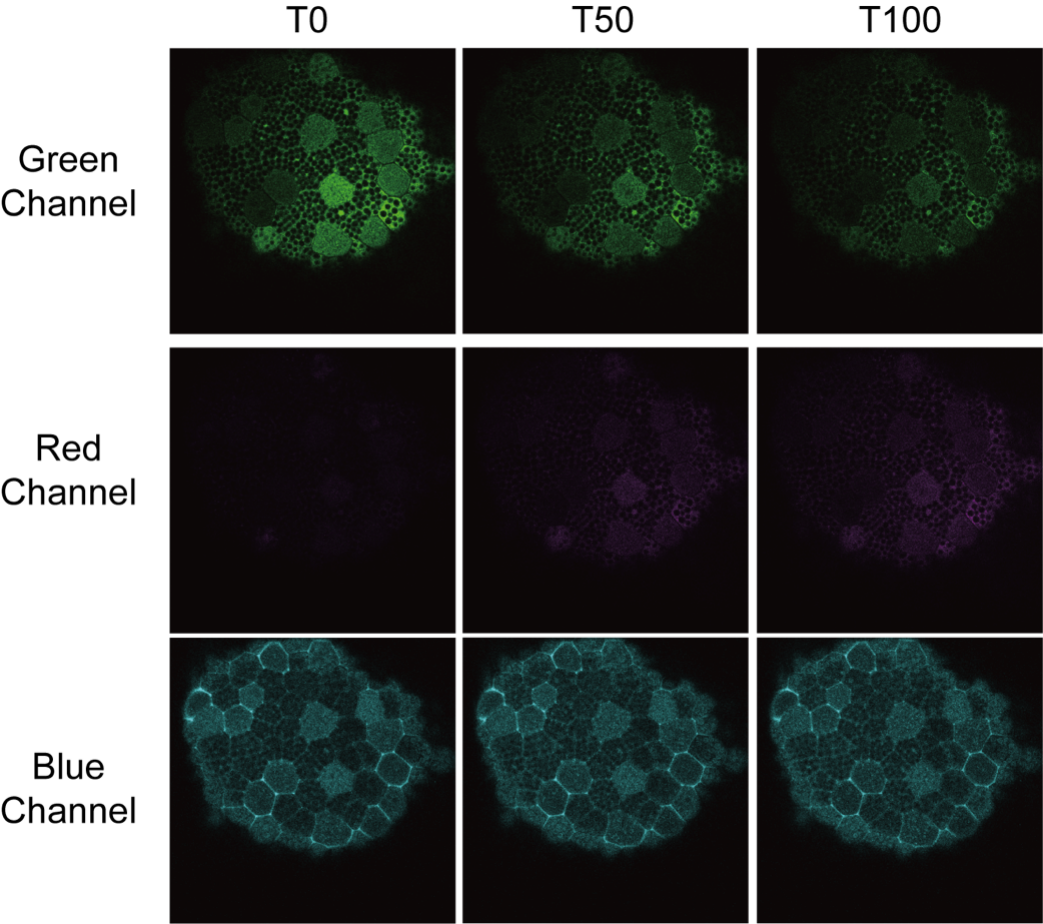
Fig 2: In Marchantia polymorpha, we observed a gradual increase of red fluorescence after the induction of conversion.
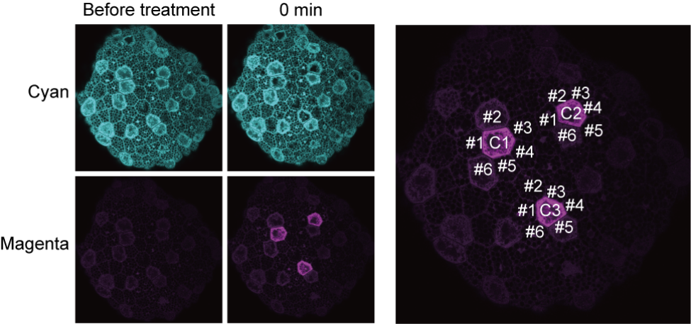
Fig 3: Single-cell photo-conversion. Photo-conversion was induced in three different Marchantia
epiderma cells. The fluorescent intensity changes in the induced cell and the surrounding cells were
measured to determine the movement of fluorescent proteins.
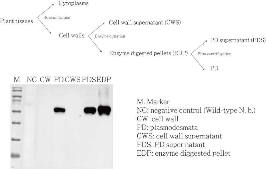
Fig 4: The PD isolation procedure. We observed signals of our target protein in the western blot analysis only after the cell wall digestion.
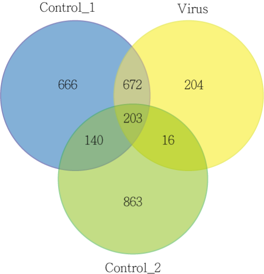
Fig 5: Protein identified by mass spectrometry after biotin labeling. We will survey through the virus only genes for further analysis.